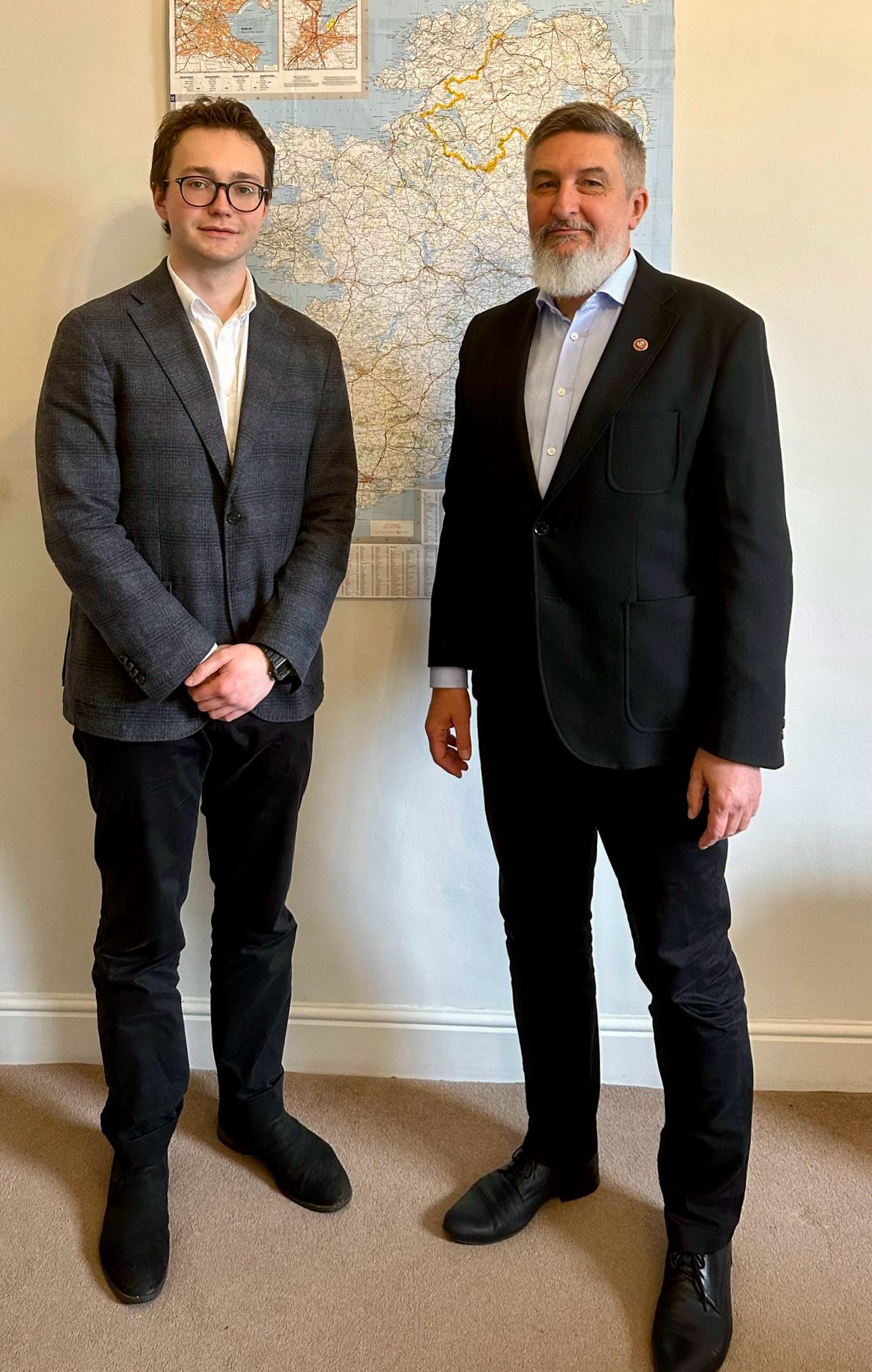
Trinity’s Dr Paula Murphy, from the School of Natural Sciences, has discovered why foetuses move in the womb.
Murphy’s research found that for biological signals – crucial for the development of bone and cartilage – embryonic movement is required. Molecular interactions, stimulated by movement, help guide cells and tissues within embryos to form a malleable, yet strong, skeleton.
Lack of movement within the embryo has been shown to result in a loss of specific signals, which can lead to abnormalities in skeletal function, such as brittle bones.
Murphy co-led the research, along with a group from the Indian Institute of Technology in Kanpur. In a press statement, Murphy said: “The relative lack of understanding around how cartilage was directed presented an unfortunate knowledge gap because there are many painful, debilitating diseases that affect joints, like osteoarthritis, and because we also often injure our joints, which leads to them losing this protective cartilage cover. Our new findings show that in the absence of embryonic movement the cells that should form articular cartilage receive incorrect molecular signals, where one type of signal is lost while another inappropriate signal is activated in its place.”
“In short, the cells receive the signal that says ‘make bone’ when they should receive the signal that says ‘make cartilage’”, she said.
Scientists have long understood the cell signalling required to build bone but understand much less about how cartilage at a joint is directed to grow. Prior to Murphy’s discovery, scientists understood that restricting movement in chick and mouse embryos resulted in a reduction of cells at joints, which caused joints to malform or fuse together completely. However, they did not understand why.
Murphy’s discovery could be used to develop better mechanisms for correct cell development, which will stop embryonic and foetal skeletal abnormalities.


